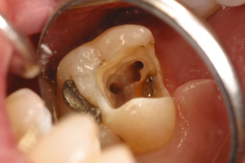
The Easy Macro View is a tool allowing dental practitioners to see the area to be treated very closely, via an endoscopic micro-camera.
Reviews
The concept
Technology
Fonctionnalities
Characteristics
Configuration
Reviews
No customer comments for the moment.
To your expectations EKLER brings a new answer. A new vision, a near vision.
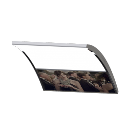
Whatever you do, you can appreciate the exceptional image quality.
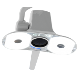
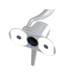
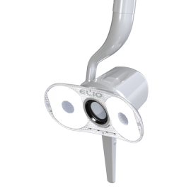
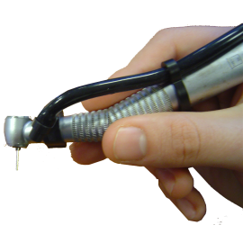
A unique solution, a mini-camera in-situ accompanies you and assists you throughout your intervention. This is the new EMV (Easy Macro View) solution.
Clipped on the rotating instrument the mini camera has a privileged location. Connected to the air system to chase moisture from the lens it offers the user images of faithful of your care.
EMV (Easy Macro View) is an unprecedented solution allowing the vision of the operative field thanks to the use of an endoscopic camera placed on your instruments.
With a diameter of 3.4mm our camera is designed to withstand operating conditions such as spray, saliva and other materials. The endoscope is enclosed in a sealed case protected by a laminar flow generating an overpressure in front of the optics. This avoids the risk of visual impairment due to liquid splashes.
To ensure better image quality we have made the choice of CMOS technology 1/10 inch. No adjustment is required. The image remains clear regardless of the position of the camera if the distance is greater than 12mm.
Images can be viewed and saved on your computer as a still image or video.
The camera is attached to the instruments with a specially designed flange. A multitude of flanges of different shapes allows the EMV to be installed on most rotating instruments.
The close vision of the surgical field is a new possibility to accompany you throughout your actions. EMV therefore becomes an important aid to the surgical decision in real time.
These applications are multiple: visual aid, traceability, communication, training ...
Visual help
EMV represents a significant contribution to dental practice. Whether you are a general practitioner or a specialist, you will benefit from this vision. The projected image near that in full screen will allow you to better appreciate the details of the field on which you intervene. The various computer tools will allow you to actually operate in augmented.
Traceability
Some acts require to be filmed backed up. EMV allows you to film your interventions and store them as your hard drives.
These records can be used either for patient follow-up and the evolution of this care.
They can also be used by your colleagues, partners, substitutes or potential purchaser.
PERFORMANCES
- Zone active : Color CMOSAnalog SquareGA™ (400x400) Image Sensor with Pixel3-HS™ Technology
- Taille du capteur: 1815 x 1815µm Analog/ DigitalAnalog.
- Correction des couleurs
- Taille de 1224 x 1212µm pour une Résolution de 400 x 400
- CSP3 Optique Format 1/10"Pixel, taille de 3.0 µm
- Affichage en temps réel de 60 images / seconde @ 400 x 200
30 images / seconde @ 400 x 400 - FullPower Consumption Standby: TBD
Active: 80 mW Température Stable: 0° - 50°C
Fonctionnel sous : -20° à 70°C
DIMENSIONS
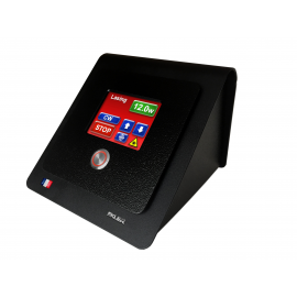
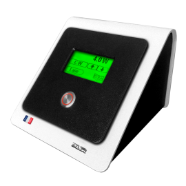
- Carte de gestion boitier de 70*105 pour intégration unit.
- Un seul cordon ressort de celle-ci pour un confort d'utilisation et de polyvalence multi-pièce à main.
- Dimensions zone active du capteur cmos 400 x 400 pixels.
- Dimensions ext Capteur endoscopique 11,5 mm x 3,4 mm
CARACTERISTIQUES ELECTRONIQUES
- Connexion USB 2.0 , Svidéo, composite.
- Alimentation via 5 Volts ou alimentation par l'unit.
- Consommation: Environ 120 mA
- Gel et mise en tampon mémoire des images par l'intermédiaire d'une pédale USB livrée
LOGICIEL
EDIS (EKLER Dental Imaging systems)
Configuration minimum requise Ordinateur compatible PC équipé :
• d’un processeur Intel Pentium IV,
• de 512 Mo de mémoire vive,
• d’une interface USB 2.0,
• d’une carte graphique 32 Mo,
• résolution du moniteur de 1024 x 768 x 24 Bits.
Système d’Exploitation Requis :
- Microsoft Windows XP Professionnel SP3,
- Microsoft Windows Vista Professionnel SP2,
- Microsoft Windows 7 Professionnel.
- Microsoft Windows 10 Professionnel
Lien vers les logiciels de gestion EDIS LINK™ inclus.
Garantie : 1 an retour atelier (France)
- ElioRecommended Products
- LuximaRecommended Products
- Recommended Products
- QuickLazeRecommended Products
- Easy Macro ViewRecommended Products
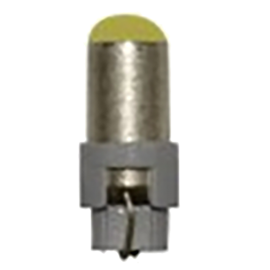
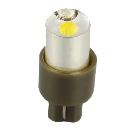
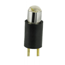
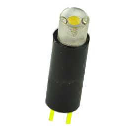
- Ampoules LEDRecommended Products